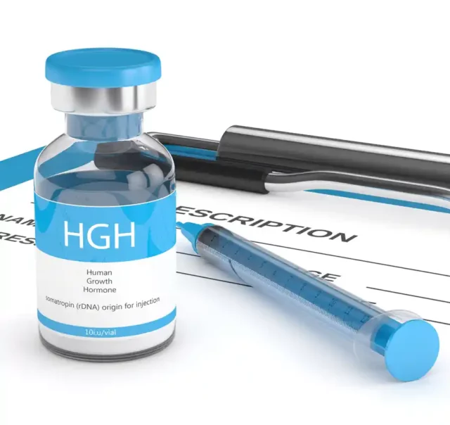


September 27, 2022 — Having soft erections and a loss of interest in sex is completely normal for men as they age. Maybe you’ve had a really stressful schedule, the bi…
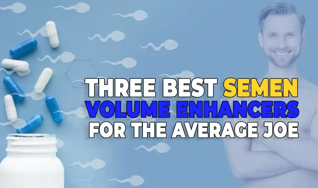
September 27, 2022 — Not every man considers the mind-blowing benefits of volume-filled orgasms, but the men who do are one step ahead. A semen volume enhancer is an a…
August 27, 2022 — Testosterone levels in men decline with age. The reduction in the volume of free testosterone levels triggers various sex health issues, including …
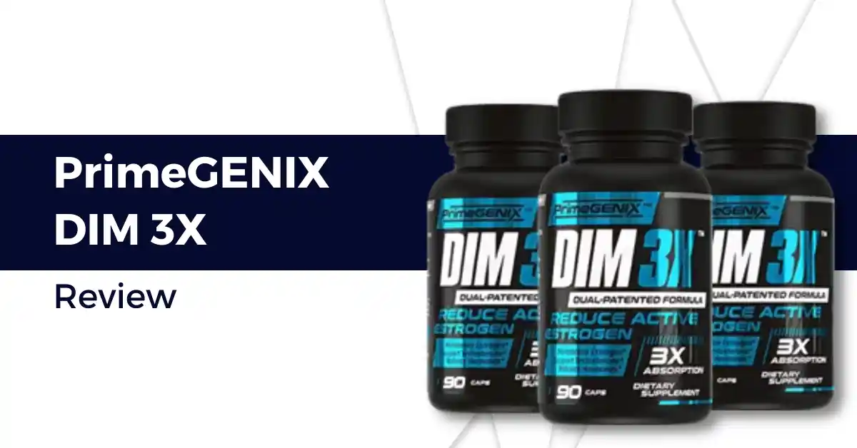
August 25, 2022 — As men get older, testosterone production decreases. At the same time, the body begins to convert more testosterone into estrogen. This transformat…
September 27, 2022 —Having soft erections and a loss of interest in sex is completely normal for men as they age. Maybe you’ve had a really stressful schedule, the big family makes it hard to get in the mood, or you’ve been with your partner for so long you’re scare…
September 27, 2022 —Not every man considers the mind-blowing benefits of volume-filled orgasms, but the men who do are one step ahead. A semen volume enhancer is an all-natural supplement that helps you achieve those never-ending orgasms. If you’re ready to ex…
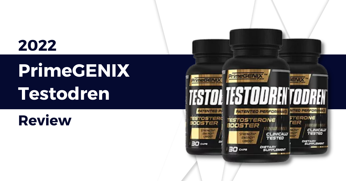
August 27, 2022 —Testosterone levels in men decline with age. The reduction in the volume of free testosterone levels triggers various sex health issues, including low semen volume, infertility, erectile dysfunction, low libido, decrease in muscle mass, and other …
August 25, 2022 —As men get older, testosterone production decreases. At the same time, the body begins to convert more testosterone into estrogen. This transformation can cause you to feel lethargic, lose muscle mass, and gain weight. Taking a supplement is a gr…
August 25, 2022 —Testosterone serves many purposes in men—from regulating your sex drive, building muscle mass, to producing red blood cells and sperm—it’s an important hormone that unfortunately declines with age. This decline pushes many men to look for ways to …
March 8, 2022 —Our hormones have far-reaching effects on human health, with imbalances affecting everything from our mental state to our body composition. If you have confirmed or suspected low testosterone levels, naturally you want to correct the problem in o…
March 6, 2022 —We can all agree that having a deficiency of testosterone affects both men’s well-being that leads to several dysfunctions. Luckily, there are prescription medications that can help men who have low testosterone. Testosterone cypionate, also…
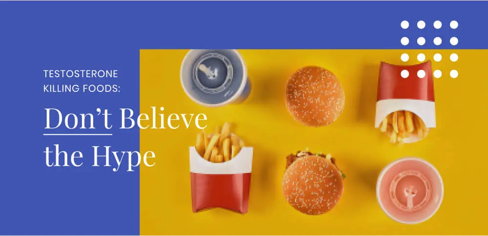
March 4, 2022 —Worrying about low testosterone has become one of the latest health panics. It doesn’t help that testosterone is, both biologically and culturally, so important to the male ideal. As a result, while it may seem overdramatic, accusing soy of killin…
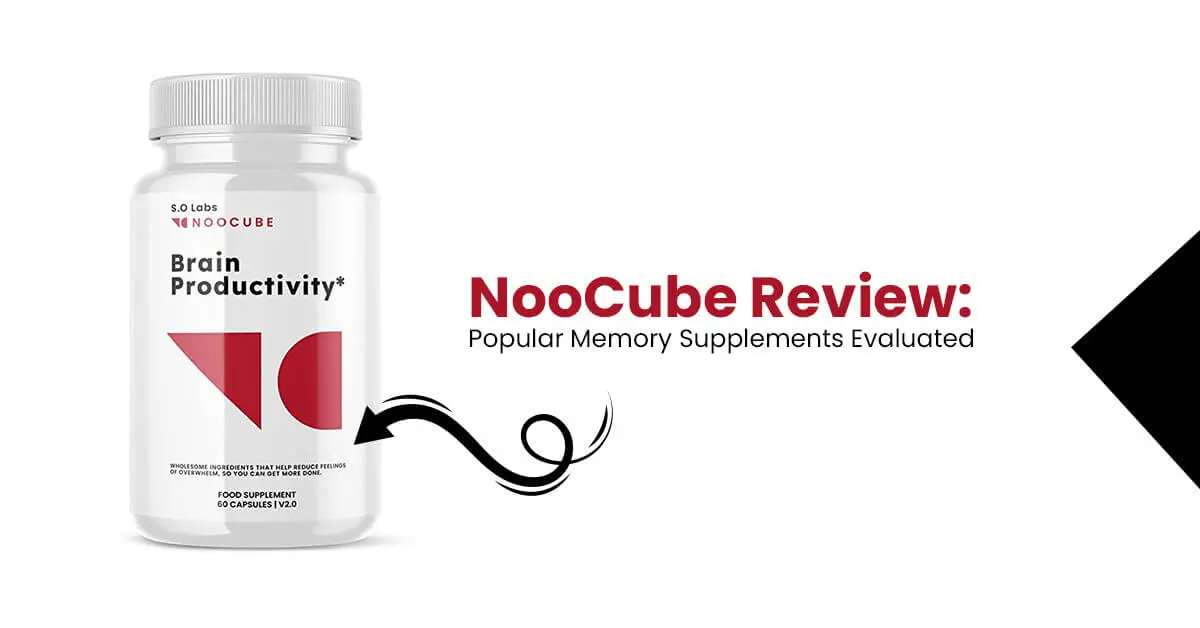
March 3, 2022 —Looking for a nootropic supplement to boost your brain function? Choosing the right nootropic isn’t easy. The market is flooded with a wide variety of products for improving cognitive function. NooCube is one of the most popular ones out t…
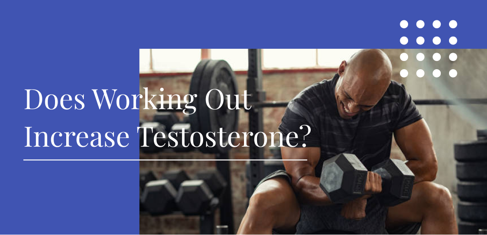
March 3, 2022 —Increasing testosterone in a natural and safe way is desired by many people. While working out can definitely do the trick, beginners can make many mistakes that set them back. They may not exercise with enough intensity, or they may overdo it. T…
September 27, 2022 — Having soft erections and a loss of interest in sex is completely normal for men as they age. Maybe you’ve had a really stressful schedule, the bi…
September 27, 2022 — Not every man considers the mind-blowing benefits of volume-filled orgasms, but the men who do are one step ahead. A semen volume enhancer is an a…
August 27, 2022 — Testosterone levels in men decline with age. The reduction in the volume of free testosterone levels triggers various sex health issues, including …
August 25, 2022 — As men get older, testosterone production decreases. At the same time, the body begins to convert more testosterone into estrogen. This transformat…